Clinical Papers & Literature
Featured Clinical Papers
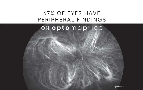
Feasibility and clinical utility of ultra-widefield indocyanine green angiography.
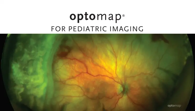
Non-contact ultra-widefield imaging of retinopathy of prematurity using the Optos dual wavelength scanning laser ophthalmoscope.
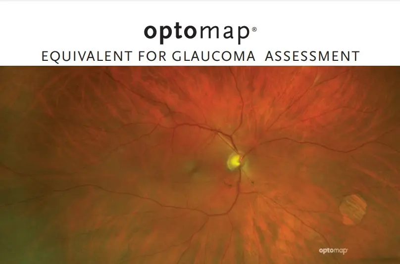
Can Ultra-widefield Retinal Imaging Replace Colour Digital Stereoscopy for Glaucoma Detection?
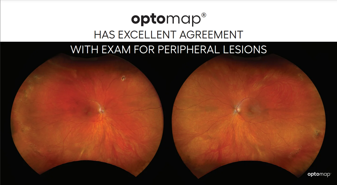
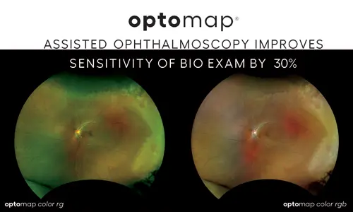
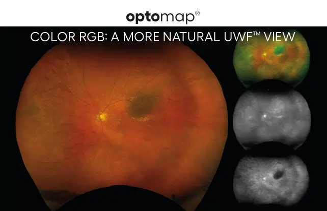
Comparison Between Ultra-Widefield Pseudocolor Imaging and Indirect Ophthalmoscopy in the Detection of Peripheral Retinal Lesions

Explore all Clinical Papers & Literature
Keyword Search
Filter by Year
Topic
Modality
Is There a Nonperfusion Threshold on OCT Angiography Associated With New Vessels Detected on Ultra-Wide-Field Imaging in Diabetic Retinopathy?
Although the NPI was significantly higher in eyes with PDR compared with severe NPDR eyes, its measurement on the whole wide-field OCTA image was not sensitive enough to replace the detection of NV for the diagnosis of PDR.
Peripheral Lattice Degeneration Imaging with Ultra-Widefield Swept-Source Optical Coherence Tomography.
UWF SS-OCT can be a useful tool to understand anatomical changes and pathophysiology of peripheral lattice degeneration.
Proliferative Sickle Cell Retinopathy in the Retinal Periphery Detected by Ultra-Widefield Imaging: A Case Report.
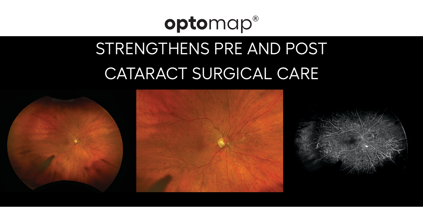
Comparison of Standard 7-Field, Clarus, and Optos Ultrawidefield Imaging Systems for Diabetic Retinopathy (COCO Study).
These findings suggest that when evaluating the 7F area on Clarus and Optos, DR severity grades are comparable to standard 7F imaging. However, it is important to understand the unique attributes and differences of each fundus camera when changing the type of system used in a clinical setting due to upgrading equipment. Additionally, if the facility has access to > 1 device, there should not be an exchange between cameras for the same patient.
New Imaging Technology for Simultaneous Multiwavelength-UWF Fundus Fluorescein Angiography and Indocyanine Green Angiography With Navigated Central and Peripheral SS-OCT.
First-in-human study of a new technology providing UWF RG/FFA/ICGA with simultaneous navigated central and peripheral SS-OCT can guide clinical management and provide new insights and understanding of central and peripheral retinal and choroidal disease.
Automatic Detection of Peripheral Retinal Lesions From Ultrawide-Field Fundus Images Using Deep Learning.
Deep Learning-Based Prediction of Axial Length Using Ultra-Widefield Fundus Photography.
Ultra-Wide-Field Fluorescein Angiography Assessment of Non-Perfusion in Patients with Diabetic Retinopathy Treated with Anti-Vascular Endothelial Growth Factor Therapy.
Qualitative and quantitative evaluation of diabetic choroidopathy using ultra-widefield indocyanine green angiography.