Clinical Papers & Literature
Featured Clinical Papers
Feasibility and clinical utility of ultra-widefield indocyanine green angiography.
Non-contact ultra-widefield imaging of retinopathy of prematurity using the Optos dual wavelength scanning laser ophthalmoscope.

Results from published clinical studies suggest that optomap may play an essential role in glaucoma management.
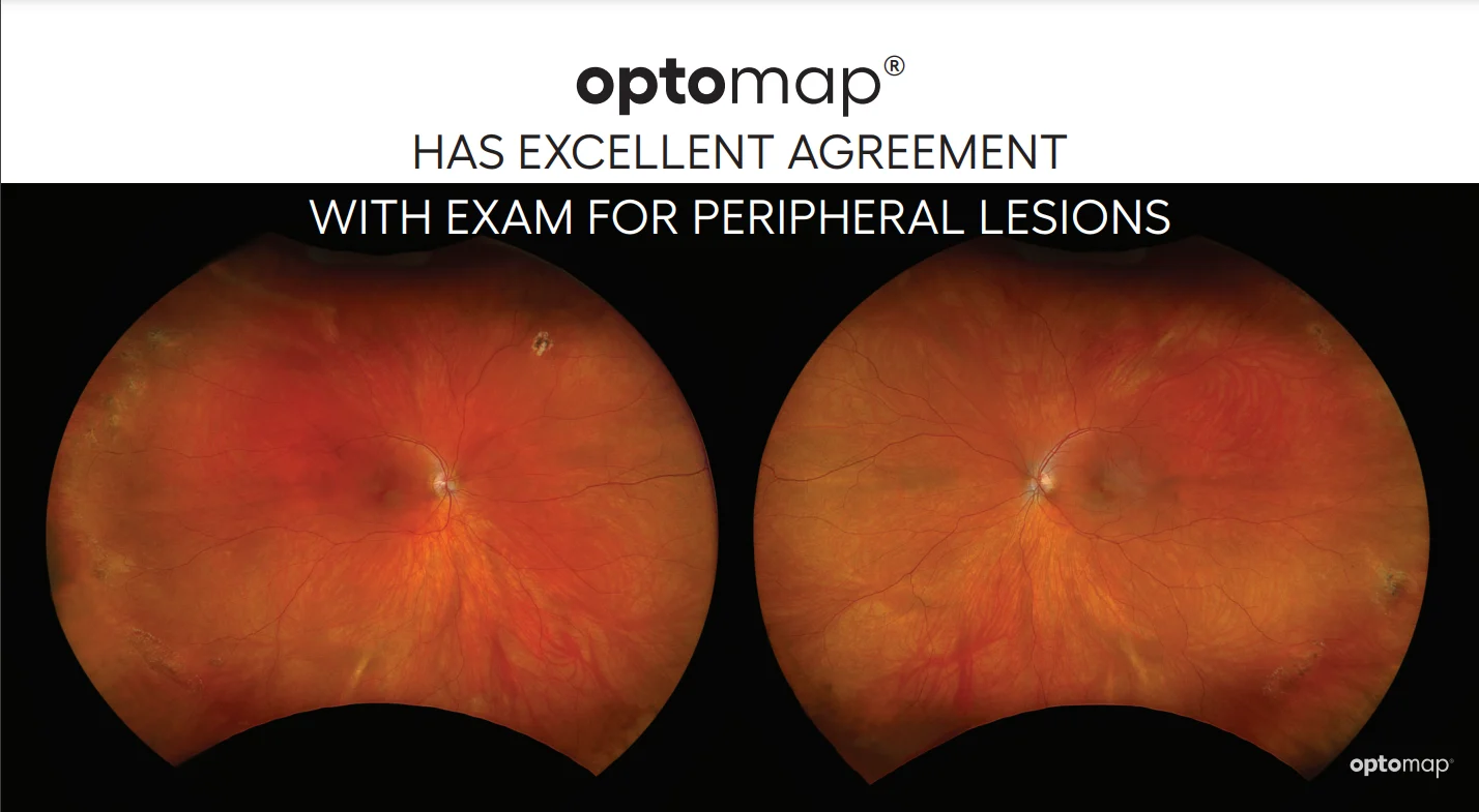
Comparison Between Ultra-Widefield Pseudocolor Imaging and Indirect Ophthalmoscopy in the Detection of Peripheral Retinal Lesions

Explore all Clinical Papers & Literature
Keyword Search
Filter by Year
Topic
Modality
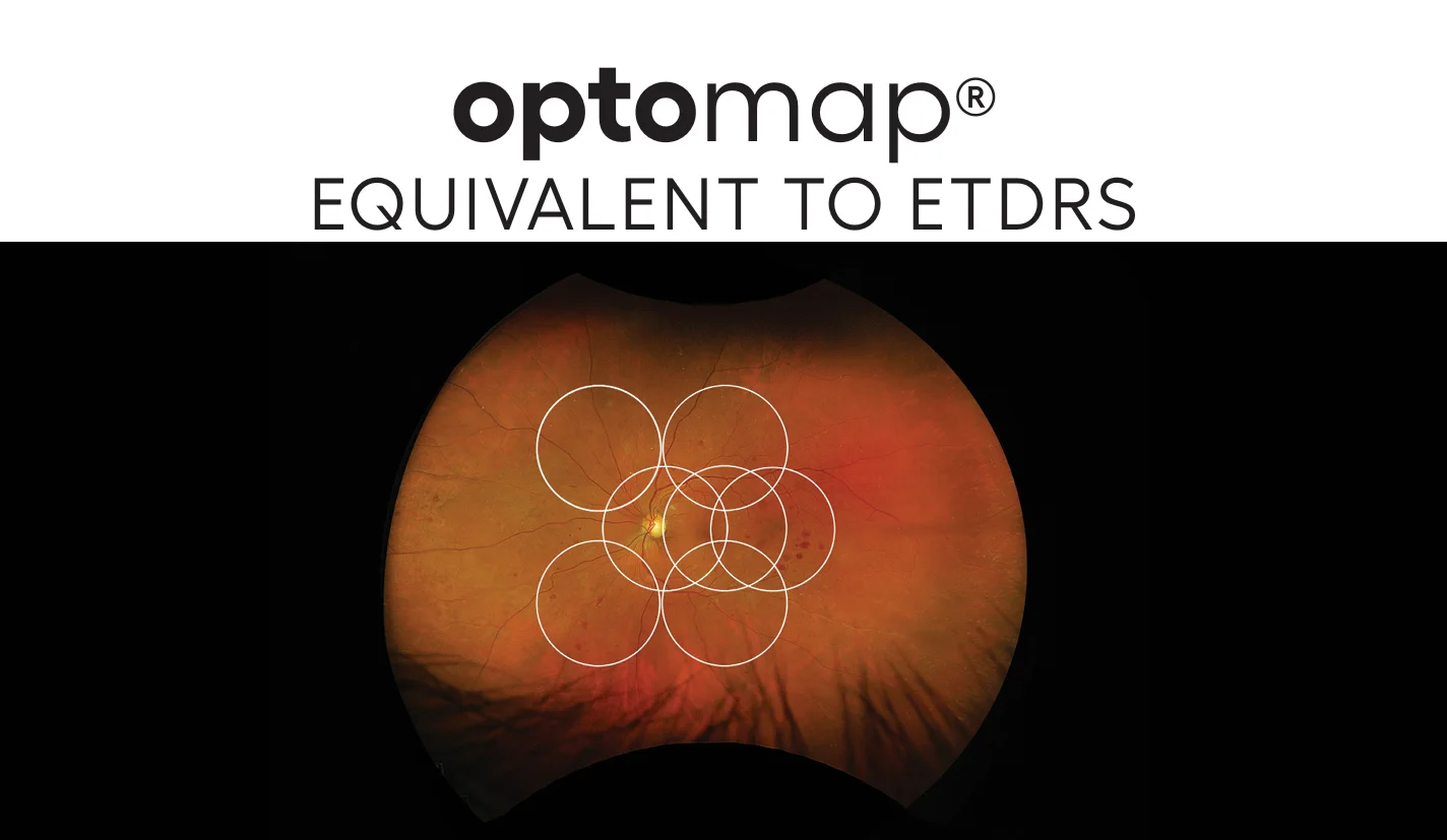
optomap Equivalent to ETDRS
Results from a large multi-center collaborative study confirm the equivalence of optomap to ETDRS Gold Standard for grading diabetic retinopathy.
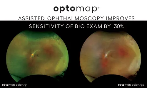
Comparison of Image-Assisted Versus Traditional Fundus Examination
Image-assisted fundus examination may enhance detection of retinal lesions by up to 30% compared with traditional fundus examination alone.
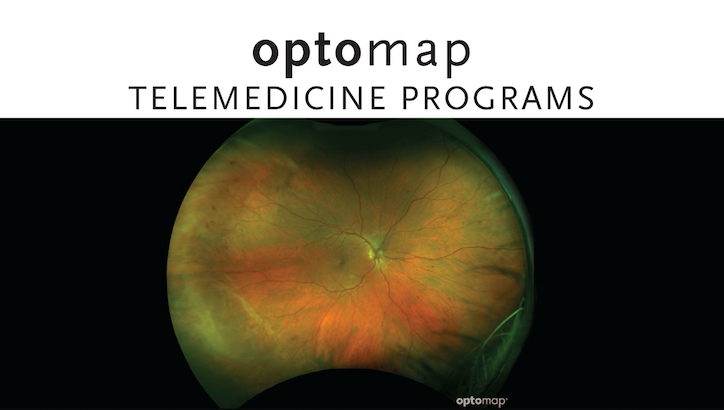
optomap Helpful in Telemedicine Programs
Several studies published in Diabetes Care have found that optomap detected 17% more diabetic retinopathy and was more efficient than traditional non-mydriatic fundus imaging.
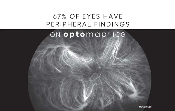
Feasibility and clinical utility of ultra-widefield indocyanine green angiography.
optomap icg images were comparable in the central pole to standard imaging devices and peripheral changes were visualized outside of 60° field of view in 67% of eyes.
Non-contact ultra-widefield imaging of retinopathy of prematurity using the Optos dual wavelength scanning laser ophthalmoscope.
The Optos ultra-widefield scanning laser ophthalmoscope is capable of acquiring clinically useful high-quality images of the fundus in ROP subjects. The imaging technique could potentially be used in monitoring ROP progression and documenting ROP regression following treatment.
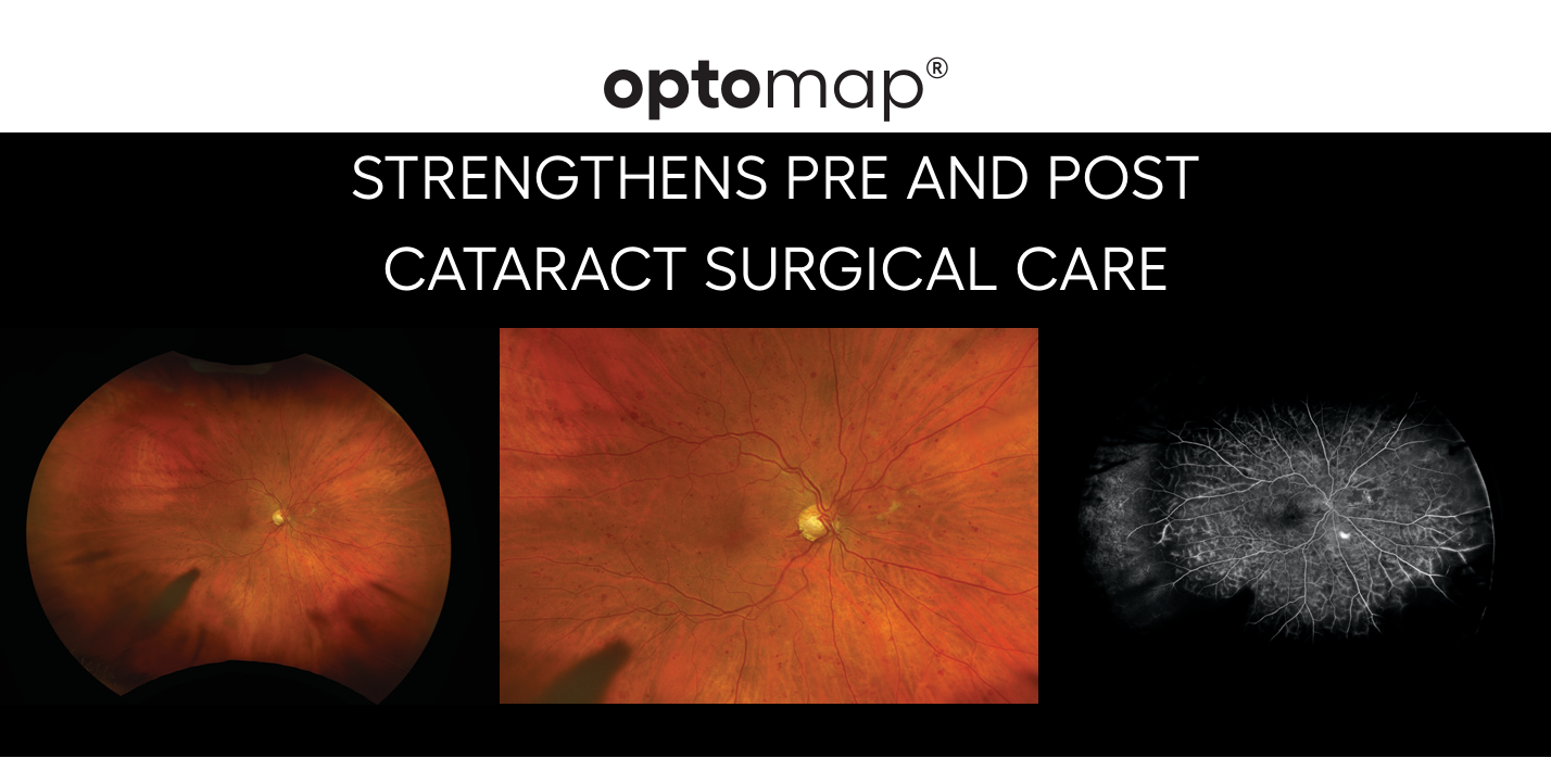
optomap Strengthens Pre and Post Cataract Surgical Care
Ultra-wide field imaging system and traditional retinal examinations for screening fundus changes after cataract surgery.
Results from published clinical studies suggest that optomap may play an essential role in glaucoma management.
Comparison Between Ultra-Widefield Pseudocolor Imaging and Indirect Ophthalmoscopy in the Detection of Peripheral Retinal Lesions
The purpose of this study was to compare the agreement for indirect ophthalmoscopy and ultra-widefield (UWF™) imaging in detecting peripheral retinal lesions predisposing to retinal rhegmatogenous detachment.
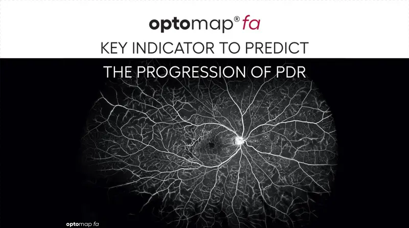
optomap fa - Key Indicator in Progression to PDR
Research using optomap fa reveals that 50% of eyes with baseline predominantly peripheral lesions (PPL) are at high risk for diabetic retinopathy (DR) progression.
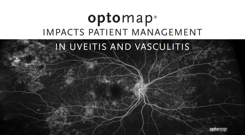
48% of Uveitis Patient's Treatment Changed with optomap
The decision to alter management was made 48% with ultra-widefield imaging compared with examination and simulated conventional FA alone.