optomap® Recognizing Pathology
This material is designed as a searchable reference resource to support clinical decision-making. The information contained here should be used as general guidance when viewing optomap and OCT images from Optos devices. The differential diagnosis should be made under the direction of the responsible physician. These images were taken on the latest ultra-widefield optomap devices.
Flap or Horseshoe Retinal Tear
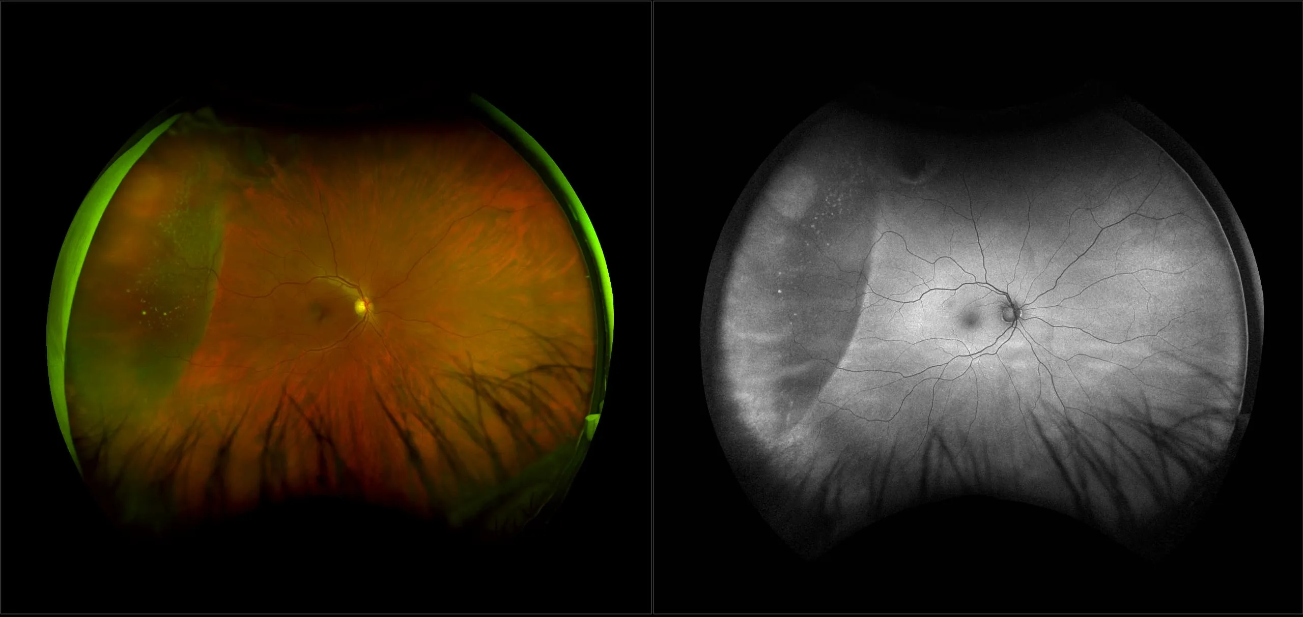
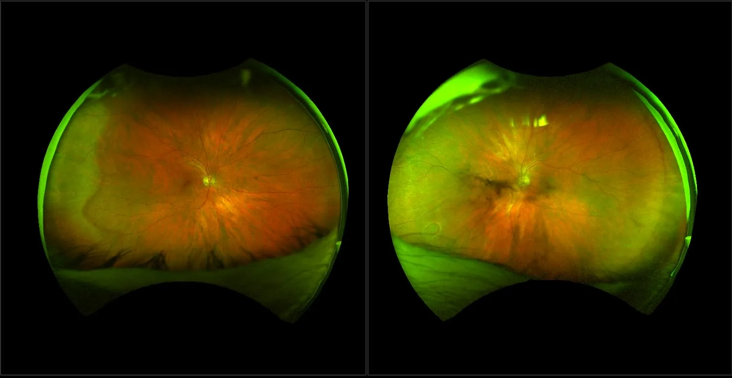
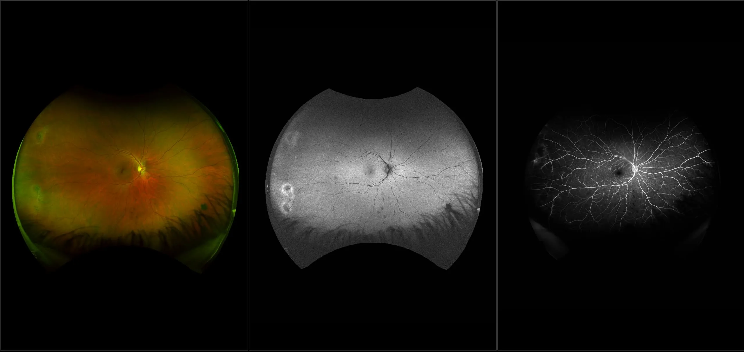
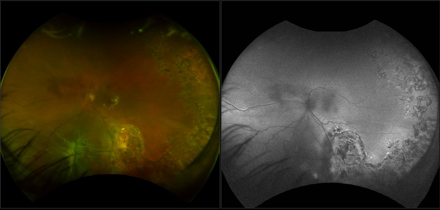
A flap (horseshoe) tear results from vitreous traction that pulls a tear of sensory retina that almost always remains attached at the anterior margin of the break. However, occasionally, the flap may be pulled free (avulsed) and be seen as an elongated operculum floating above the break. The tearing of retina usually lifts the retina at the margins and thus, can produce white margins (sometimes known as wet margins). The most frequent cause of a flap tear is a PVD. The appearance is of a horseshoe-shaped red break with a flap pulled up into the vitreous cavity. The separated posterior vitreous cortex is usually attached to it (usually can’t be seen on ophthalmoscopy). Retinal breaks are best seen with the green laser separation. Flap tears are most often found between the ora serrata and the equator where the retina is thinner than in the posterior region. Because vitreous traction may have been pulling on the site (increased vitreoretinal adhesion) for months to years before the formation of a tear, this physical traction may cause reactive RPE hyperplasia in the retina. Therefore, pigment clumping may be present at the base of the tear or on the flap. Vitreous traction usually remains attached to the apex of the flap and thus, the chances of a detachment are significant. Sometimes there may be a white collar around the break that represents a localized detachment (less than 1 DD from the edge of the break). Symptomatic flap tears have been reported to have a 25% -90% incidence of progressing to a detachment. Therefore, essentially all flap tears are treated.


