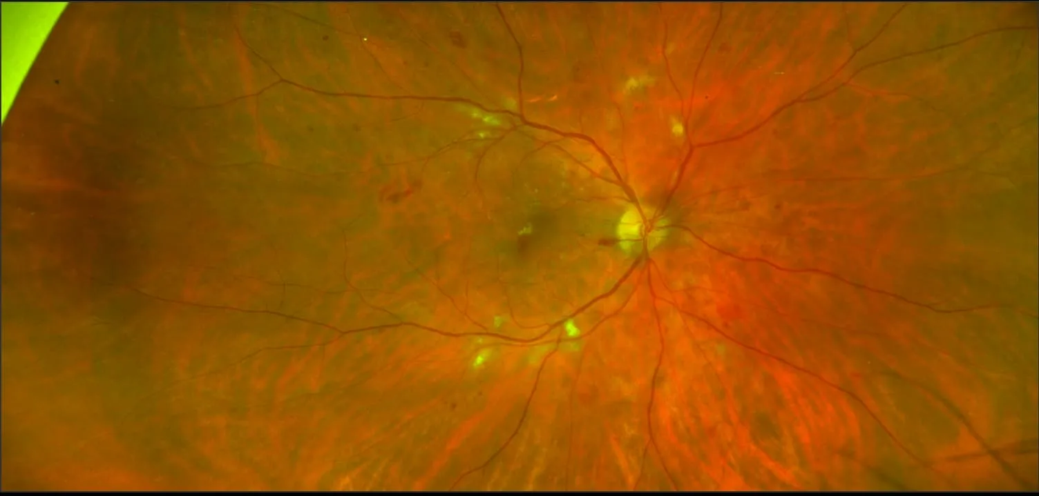
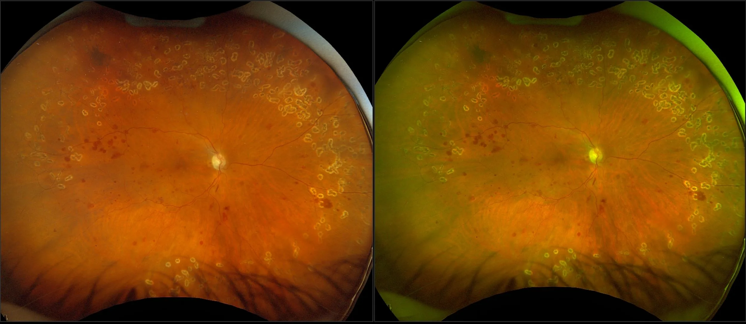
MonacoPro - Non-Proliferative Diabetic Retinopathy with PPL, RG, OCT
What: Diabetic Retinopathy is a series of progressive retinal changes that may lead to blindness that can result from long-standing diabetes mellitus. Early stage DR is non-proliferative (NPDR). It may advance to proliferative diabetic retinopathy (PDR).
Why: Optos UWF imaging has been validated as equivalent to gold standard ETDRS 7 Standard Field (ETDRS) imaging for the grading of diabetic retinopathy, in addition, lesions have been found in up to 50% of patients outside of the area captured by ETDRS. As such, UWF imaging is becoming a standard of care for imaging in the management of diabetic retinopathy.
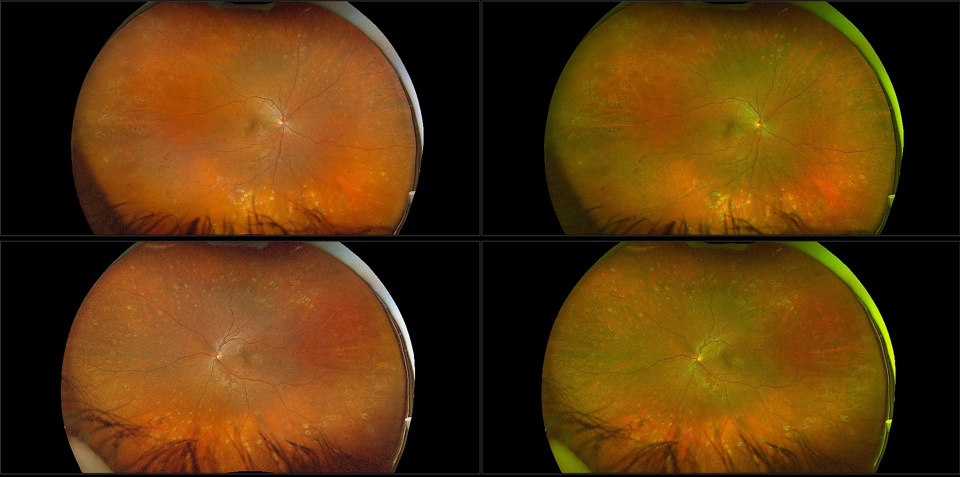
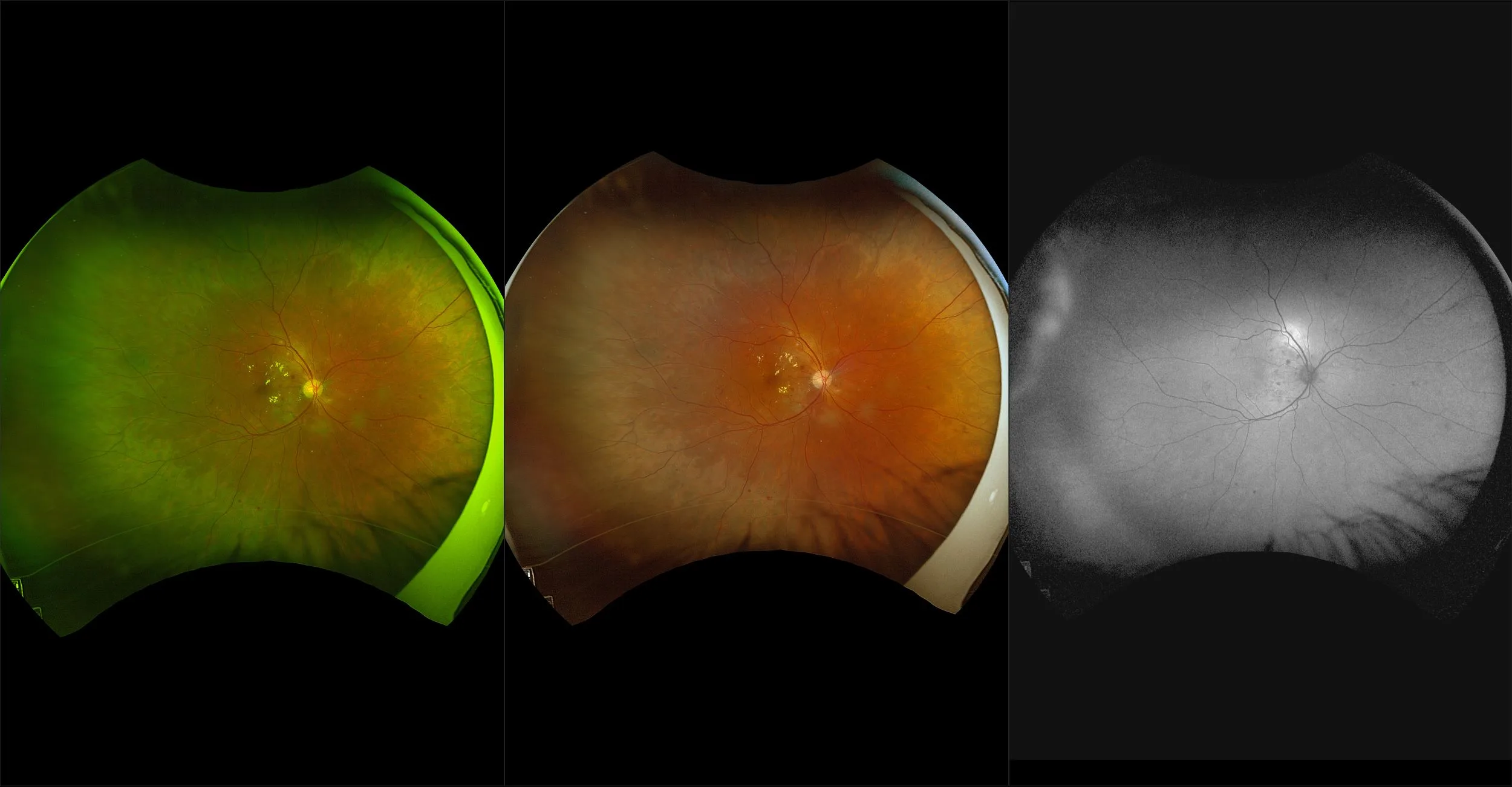
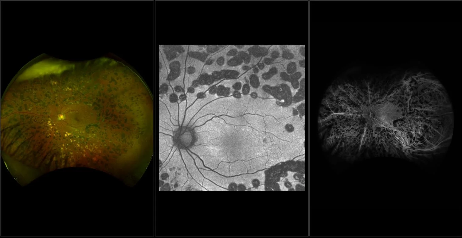
In the Picture: optomap color rg shows moderate NPDR with predominately peripheral lesions and a normal OCT. The green channel (532nm) helps to visualize the hemorrhages. Although there were no findings on the OCT, the periphery still shows lesions. One study showed that these predominately peripheral lesions (PPL), were 4x more likely to progress to proliferative diabetic retinopathy over 4 years. This shows the importance of imaging in the periphery for patients with diabetes, as this patient is likely at high risk for progression. Capturing and documenting the peripheral lesions helps determine management and treatment of diabetic retinopathy patients without compromising quality.
References:
1: Nonmydriatic Ultrawide Field Retinal Imaging Compared with Dilated Standard 7-Field 35-mm Photography and Retinal Specialist Examination for Evaluation of Diabetic Retinopathy. Silva, Cavallerano, Sun, noble, Aiello, Aiello. American Journal of Ophthalmology, 2012
2: Peripheral Lesions Identified by Mydriatic Ultrawide Field Imaging: Distribution and Potential Impact on Diabetic Retinopathy Severity. Silva, Cavallerano, Sun, Soliman, Aiello, Aiello. Ophthalmology, 2013
- Silva et al Peripheral Lesions Identified on Ultrawide Field Imaging Predict Increased Risk of Diabetic Retinopathy Progression over 4 Years. Ophthalmology, 2015

