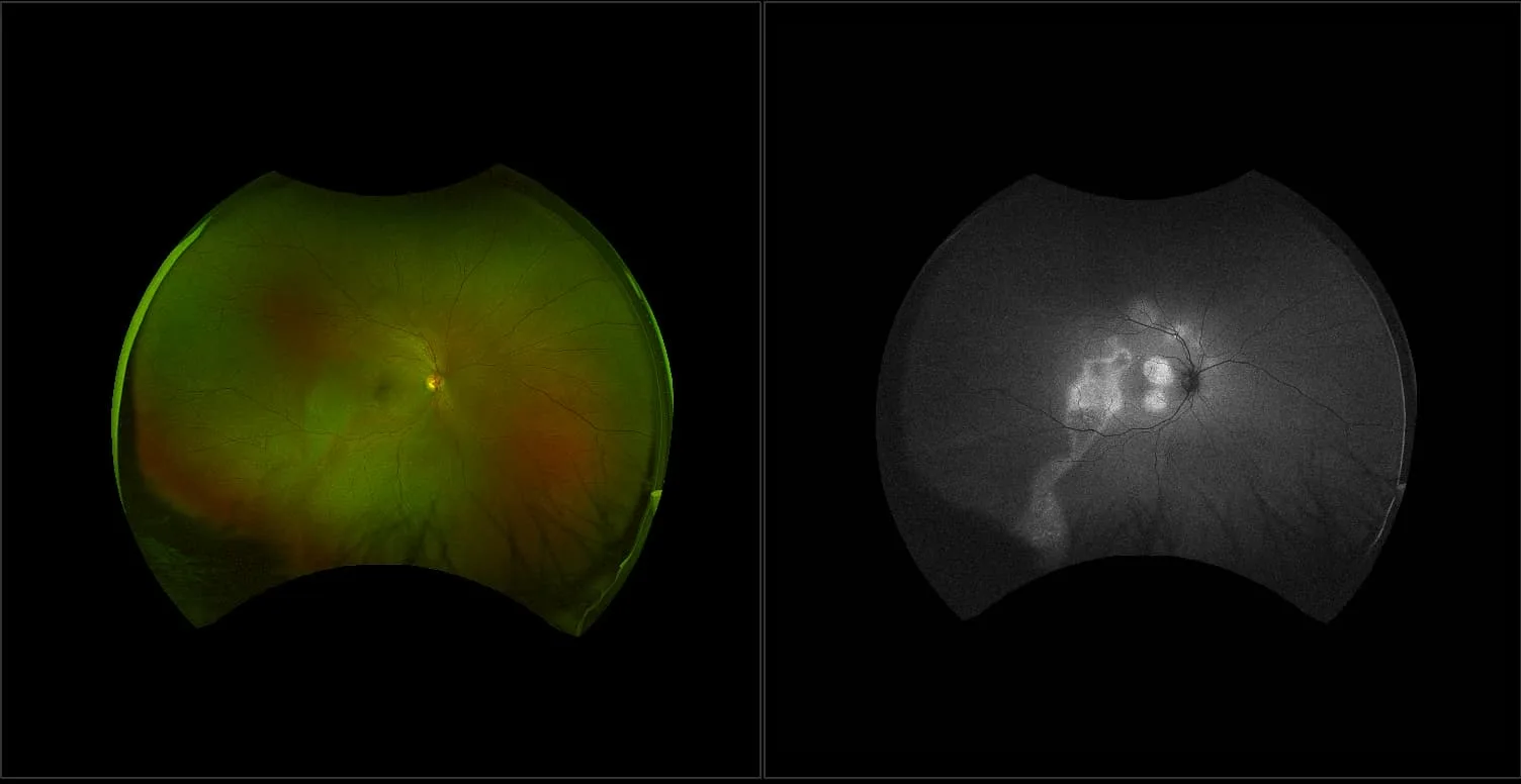
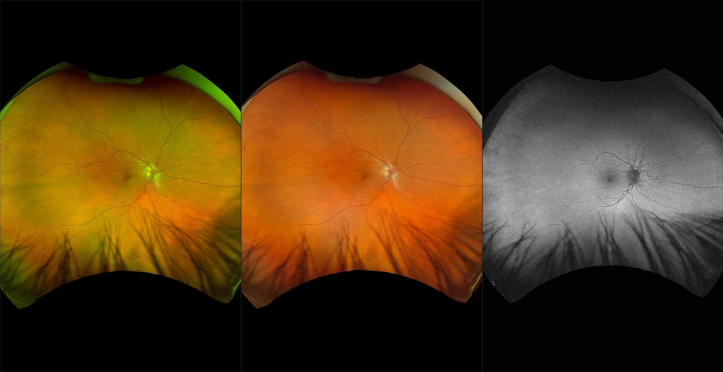
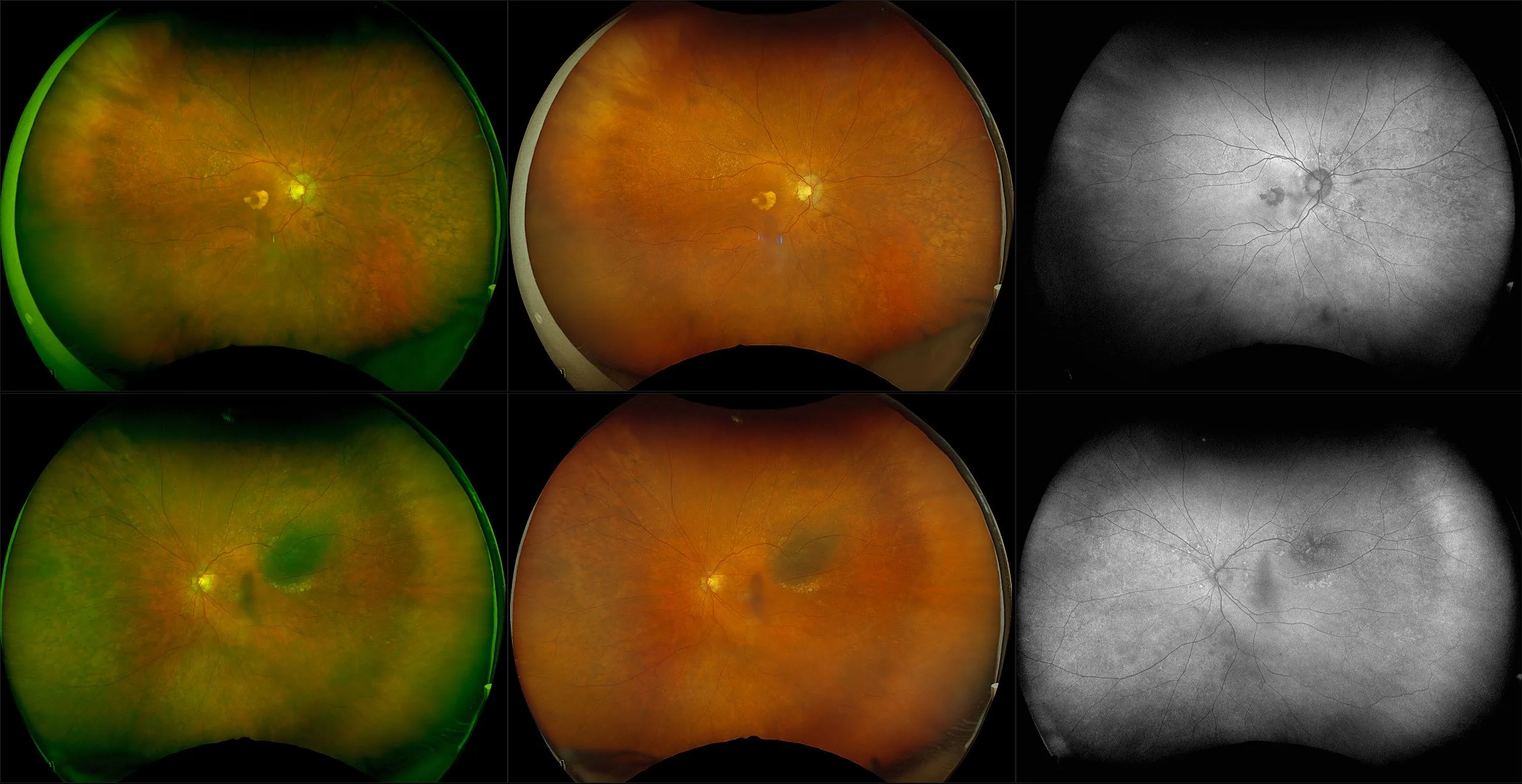
California - Central Serous Chorioretinopathy, RG, AF
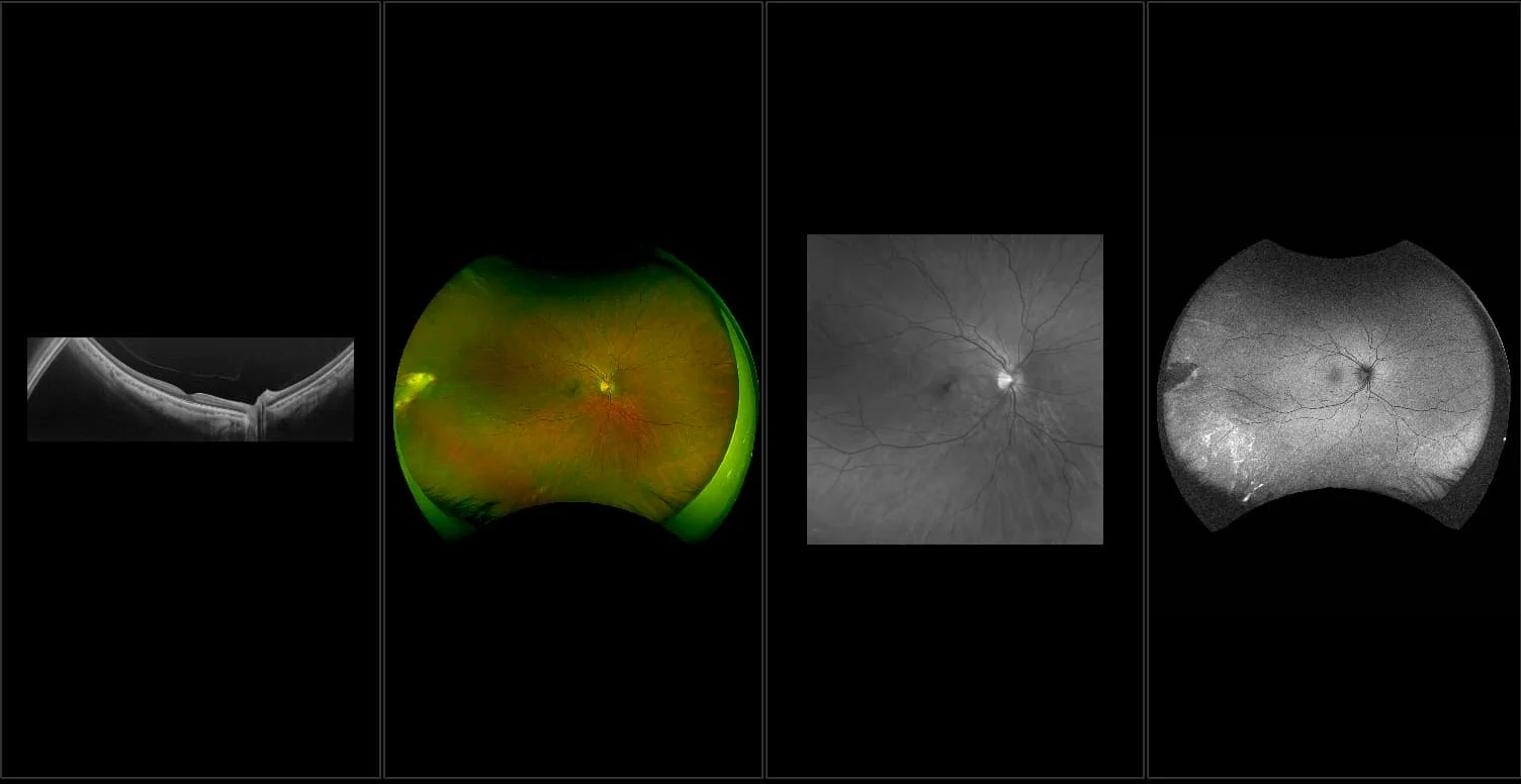
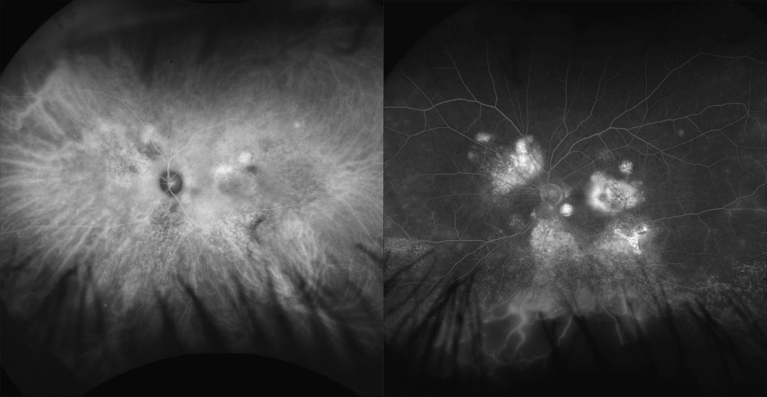
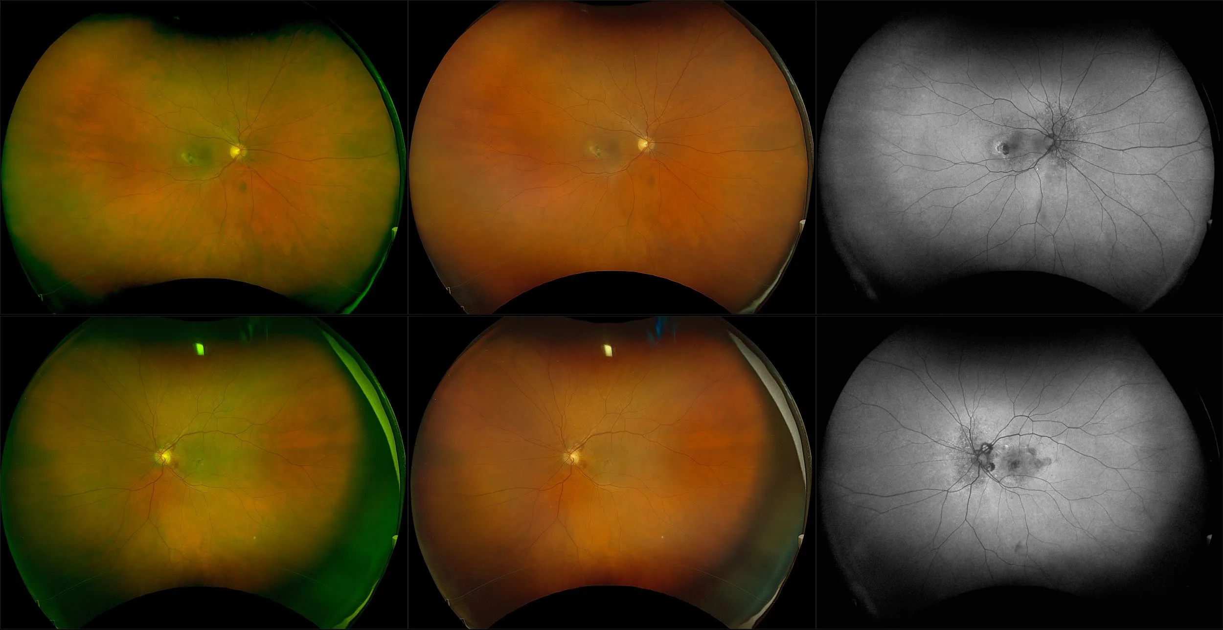
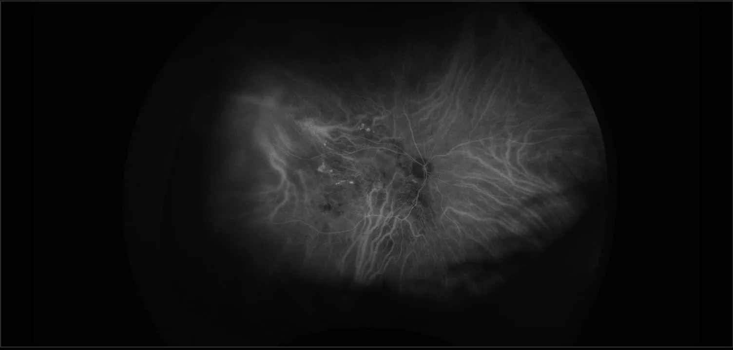
What: Central Serous Retinopathy (CSR) is a blister-like elevation of sensory retina in the macula with a localized detachment from the pigment epithelium. This results in reduction and/or distortion of vision that usually recovers within a few months.
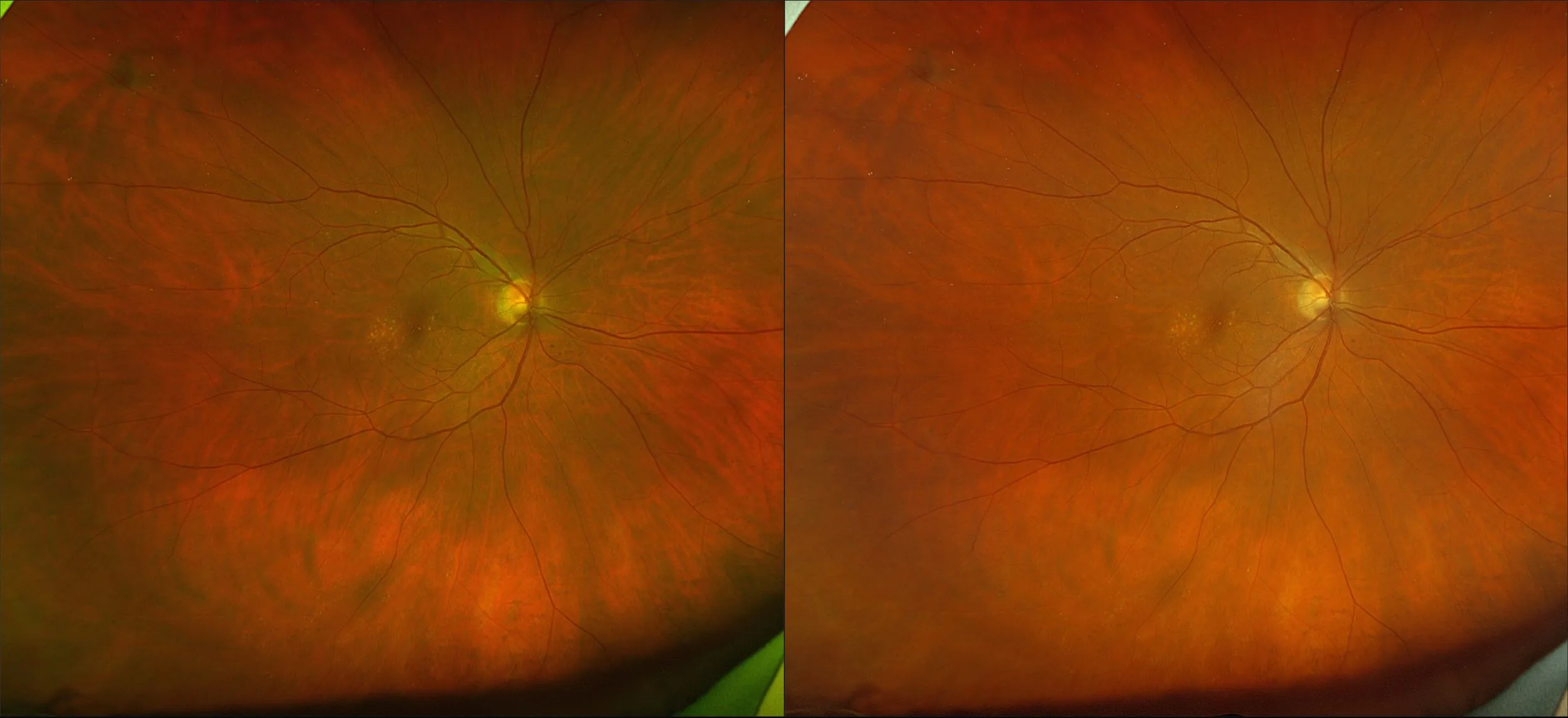
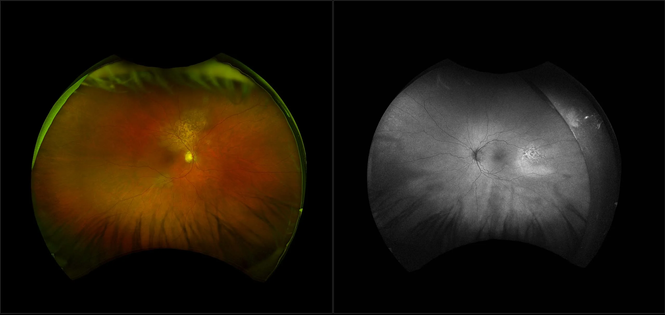
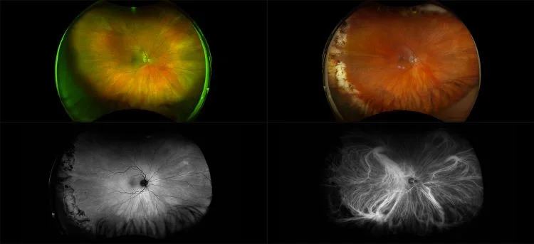
Why: optomap color images show subtle fluid build-up and macular changes in the posterior pole extending down to the inferior retina. Traditional limited-field imaging would require multiple images to capture the extent of fluid build-up in this case. One study found that nearly 52% of CSR patients have changes that extend into the periphery on color and AF imaging demonstrating the importance of capturing as much of the retina as possible.
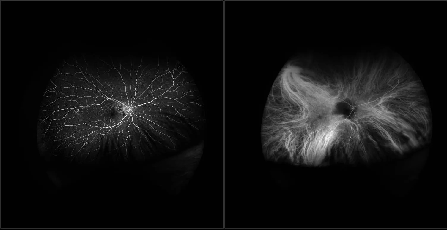
In the picture: A corresponding optomap af may show a gutter-like appearance extending to the mid-to-far periphery which is characteristic of chronic central serous retinopathy. The hypoautofluorescence gutter-like appearance corresponds to the loss of photoreceptors. Hyperautofluorescence around the edges indicates fluid accumulation and disease progression.
Reference:
- Sadda et al. Prevalence of Peripheral Abnormalities on Ultra-widefield Greenlight (532nm) Autofluorescence Imaging at a Tertiary Care Center. IOVS. 2012.
