Utilizing Autofluorescence Imaging in Your Clinical Setting
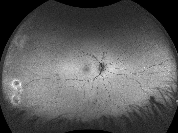
Autofluorescence retinal imaging is a non-invasive study of the fluorescent properties of lipofuscin in the RPE and provides clinical information not available using conventional imaging techniques. optomap af allows for visualization of the metabolic changes at the level of the RPE and helps to identify areas that may be at high risk. Autofluorescence imaging is a vital feature in monitoring and treating pathology in the retina.
Below we have provided some educational and diagnostic resources to help you in making the most of optomap af in your practice and clinical setting.
Webinars
Ultra-widefield Autofluorescence Imaging: A Game Changer
Mr. Simon Browning - UK
The Importance of FAF in Retinal Pathology Diagnosis
Jerome Sherman, OD
Best Practices for Utilizing UWF and FAF
Maurice Wilson, OD
Clinical Papers and Literature
optomap af Diagnostic and Patient Educational Tools
Single Page optomap af Atlas and Full optomap af Interactive Booklet

Download our optomap af Diagnostic Atlas. This tool is helpful for patient education regarding types of disease and pathology that can occur on the retina.

We have expanded on our popular retinal atlas (to the left) and created a full handbook. Each pathology has expandable images and additional educational information.
Recognizing Pathology
optomap Recognizing Pathology is designed for eye care professionals as a searchable reference resource to support clinical decision-making.
Stocked with over 200 cases and all ultra-widefield optomap image modalities plus integrated OCT.
Optos Blog
Our blog is a great resource on the benefits of optomap af imaging in your practice and clinical setting. To get you started:
Utilizing Autofluorescence in your Clinical Eyecare Setting
AMD: More Than a Macula Condition
Multiple UWF Modalities Enable More Comprehensive Peripheral Evaluation
What your Peers Are Saying

David Way, OD
Spring Klein Vision Center
Texas, USA
Morven Campbell
Iris Blue Optical
Glasgow, UK
Looking for something specific?
Let us know how we can help by completing the form below!