Optos UWF Retinal Imaging Boosts Practice Confidence
Dr. Jon Laudadio is an optometrist with two locations in British Columbia, Canada. One location houses an Optos California ultra-widefield (UWFTM) retinal imaging device, and the other location uses a traditional wide field camera. He has witnessed the substantial benefits and assurance that optomap® UWF imaging provides both him and his staff.
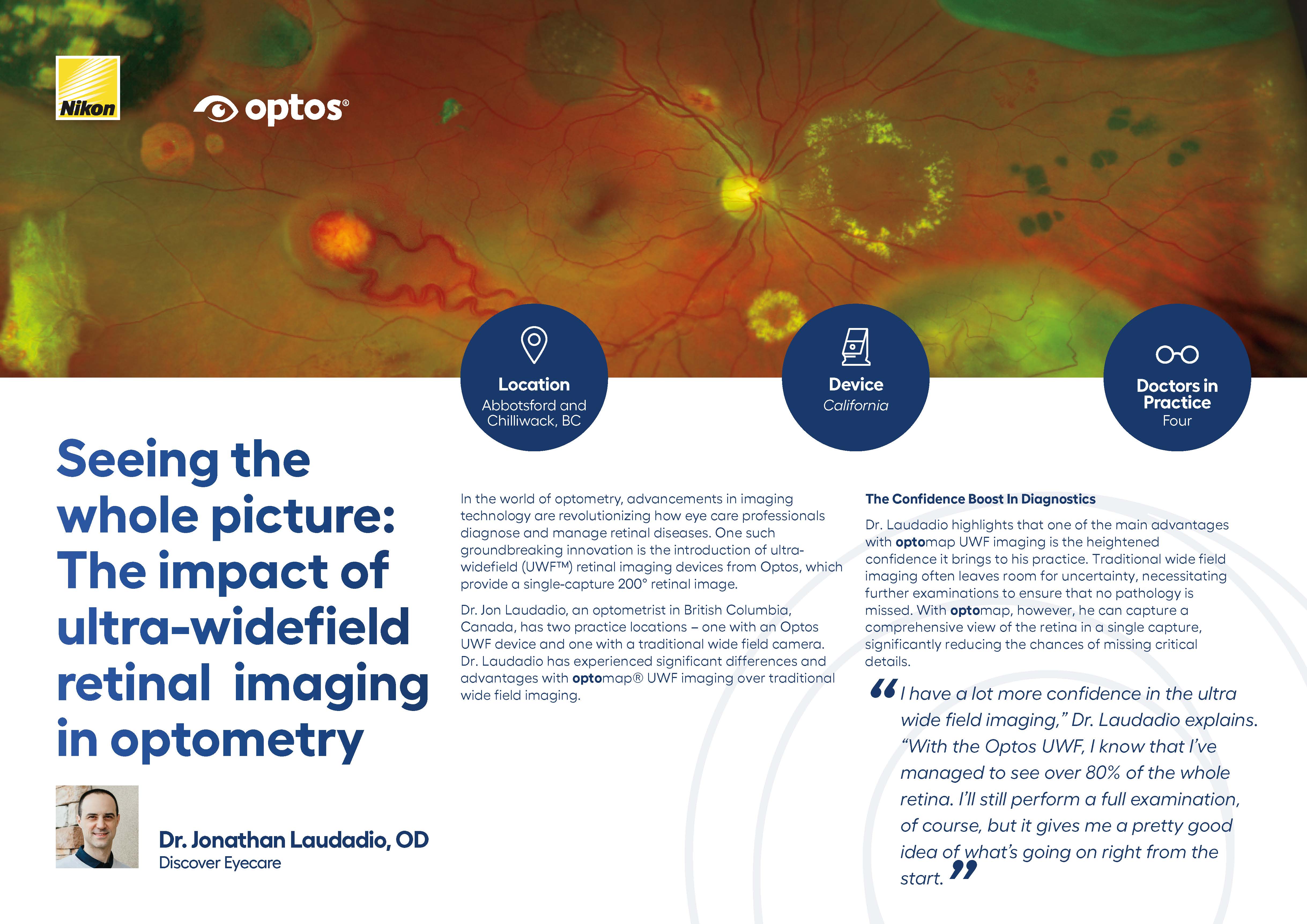
"I have a lot more confidence in the UWF field imaging," Dr. Laudadio explained. "With the Optos UWF, I know that I've managed to see over 80% of the whole retina. I’ll still perform a full examination, of course, but it gives me a pretty good idea of what’s going on right from the start."
An optomap produces a high resolution, 200° retinal image taken in a single capture. To match that field of view with traditional equipment, multiple images and in some cases, dilation is required. optomap technology reduces the amount of time a patient spends in the exam chair, eliminates the need for multiple flashes in each eye, and increases patient satisfaction.
optomap imaging is also preferred amoung technicians and staff, increasing their screening efficiency. "The staff definitely find it more irritating to take multiple images," Dr. Laudadio stated. “As soon as you take a picture with a bright flash, the patient's pupil goes small. So when you go to take your secondary wide field image, it’s a bit tougher because their pupils are so much smaller. When you just have one image on one side and one image on the other with optomap, it's so much easier for both the staff and the patient. With Optos technology it's one image per eye, making the process much quicker and more pleasant for the patient," he added.
Optos technology delivers nine imaging modalities across all devices, a larger field of view in less time, and over 2,500 clinical and peer-reviewed studies which span 235 disease states, demonstrating optomap and clinical utility go hand in hand. From diabetic retinopathy and uveitis glaucoma to tumors and pediatric vitreoretinal disorders, many pathology types identified can be treated if detected early.
Dr. Laudadio recalled one patient who complained about increased floaters and lightening flashes. Even after multiple images using the traditional wide field camera, he was still unable to diagnose the problem.
“I could visually see something was off, but I couldn’t image it with my wide field machine. I took so many pictures in all kinds of directions, but I just could not see the problem on the image. It took one image with the optomap, and there it was — a tear in the retina. It's cases like these that make me wish I had UWF imaging from the start."
The quicker the diagnosis, the quicker the care plan can be developed. Optos’ enhanced OptosAdvance software enables users to not only show their patients 3D images of their eyte to better explain an disorder, but they can also instantly annotate and share images of suspicious pathologies with specialists to expedite a treatment schedule.
Dr. Laudadio concludes. "If you come in for an exam, wouldn't you want me to have the tools that will make sure I can see as much as possible? To me, that's the obvious choice."
With more than 25,000 devices installed, Optos is redefining what a standard eye exam should look like, improving patient outcomes across the globe. If you are interested in enhancing your practice, visit our website to schedule a risk-free demonstration to see the difference optomap UWF imaging can make.