Ultra-widefield Imaging Technology in Pediatric Optometry
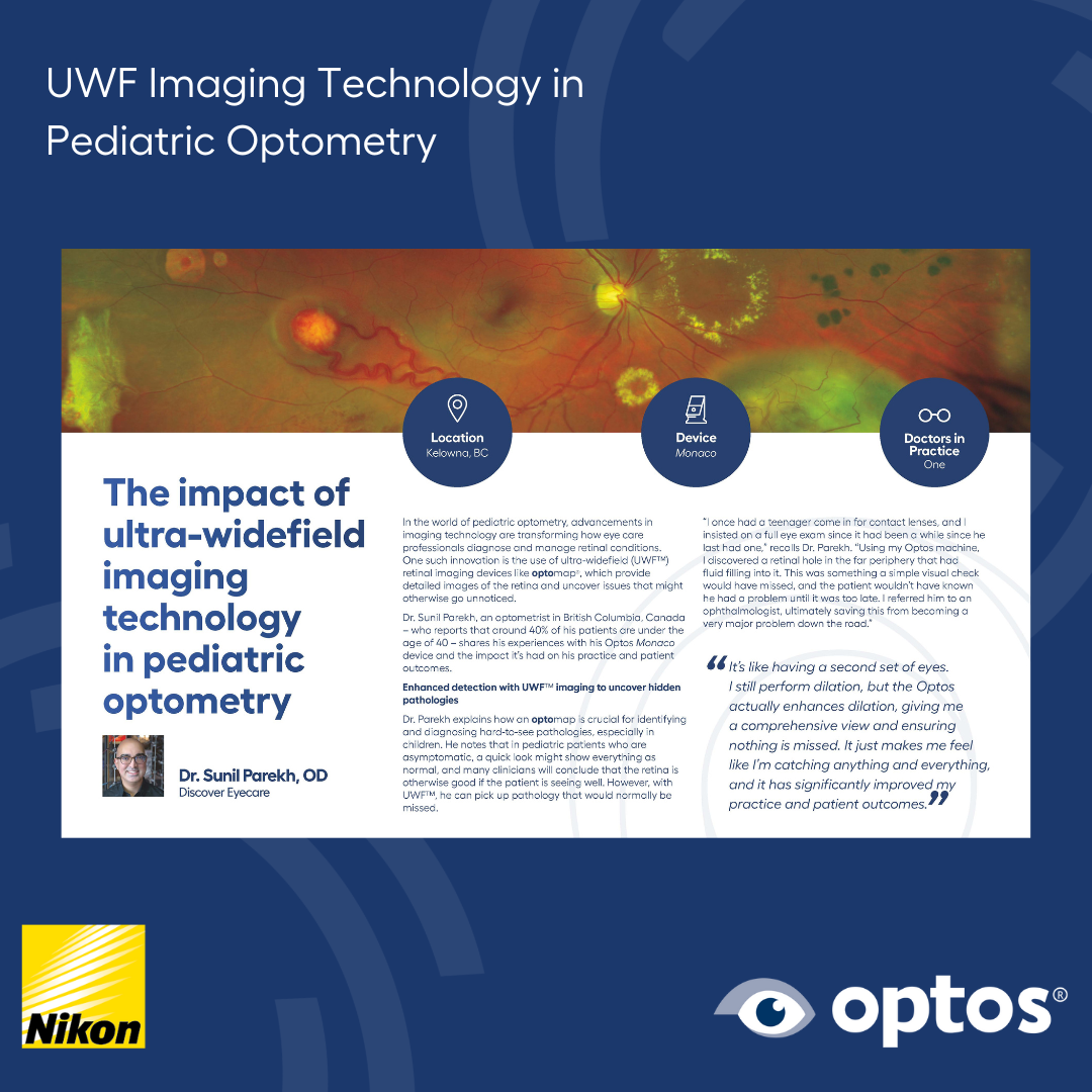
Advancements in imaging technology are renovating the standard of care within pediatric optometry practices. The use of innovative ultra-widefield (UWF™) retinal imaging devices from Optos with optomap® technology work to discover and diagnose undetected pathologies hiding in the periphery. Dr. Sunil Parekh, an optometrist in British Columbia, Canada, shared how his Optos® Monaco impacts his practice and patient outcomes.
optomap is the only technology that captures 200-degree UWF in a single shot. These images are critical, especially in pediatric patients who may have no symptoms of eye issues or vision problems.
“I once had a teenager come in for contact lenses, and I insisted on a full eye exam since it had been a while since he last had one,” recalled Dr. Parekh. “Using my Optos machine, I discovered a retinal hole in the far periphery that had fluid filling into it. This was something a simple visual check would have missed, and the patient wouldn’t have known he had a problem until it was too late. I referred him to an ophthalmologist, ultimately saving this from becoming a very major problem down the road."
Containing fundus autofluorescence (optomap af), optomap images showcase retinal structures that are invisible to the naked eye, enabling doctors to spot problems such as nerve damage that otherwise could have been missed.
"When examining the optic nerve, it might appear normal, but the autofluorescence can show abnormalities lighting up like a Christmas tree," said Dr. Parekh. “The optomap shows you so much more, giving you a significantly better understanding of what’s going on.”
Seeing is believing and with OptosAdvance™ software, patients can see the images right in the office. This provides them with a better understanding about the complexity of their eyes. Parents and children alike recognize the importance the greater field of view makes in terms of eye health.
"They start to understand the importance of comprehensive eye exams almost immediately. Many are used to quick, routine checks. They think it’s all eye charts and ‘is lens 1 better or is lens 2 better?’ But when patients get to see detailed images of their eyes, they suddenly realize the depth of the exam. It's a eureka moment for them, and they really appreciate how crucial their eye health is," explained Dr. Parekh.
Pediatric, comprehensive annual eye exams are key to early detection of eye issues, which lead to greater chances of correcting problems and preserving vision. optomap images are stored and compared year-over-year to monitor any changes or condition progression. Images are easily shared between practices for easy referrals for timely care-management plans to be created.
"Early detection is crucial. Many eye conditions are asymptomatic and can lead to severe issues like retinal detachment or amblyopia (lazy eye) if not managed early,” said Dr. Parekh. “I just take the screenshot, save it to my desktop, and send an email to an ophthalmologist with a detailed referral letter. This allows them to see precisely what’s going on. It’s really true that a picture says a thousand words!”
The diagnostic capabilities increase practice confidence and patient referrals to their friends and family.
"It's like having a second set of eyes. It just makes me feel like I’m catching anything and everything, and it has significantly improved my practice and patient outcomes. This leads to them recommending our practice to friends and family, and then the next thing I know, I'm seeing their friends and family as well! It’s had an impressive effect on increasing our patient flow."
Read Dr. Parekh's full testimonial here. If your practice is due for a transformation, or to learn about the additional benefits of implementing optomap into your practice or clinical setting, visit our website today.