A study conducted at the New England College of Optometry yielded some exciting results for practitioners using Optos’ retinal imaging devices as a part of comprehensive eye exams.
The study was designed to compare retinal imaging techniques and specifically evaluated how an optomap-assisted fundus exam is able to detect retinal lesions in comparison with traditional ophthalmoscopy. Researchers studied 339 eyes to see whether or not Optos’ ultra-widefield retinal imaging technology could allow practitioners to perform a more comprehensive exam, as the accuracy of traditional dilated retinal exams currently performed vary from 32% to 82%.
With the results recently published in Eye and Brain, the study is believed to be the first that demonstrates the ability of digital technology imaging to enhance a traditional dilated fundus exam. The study revealed that the optomap-assisted technique revealed about 30% more retinal lesions than traditional ophthalmoscopy. Researchers also concluded that optomap image assisted fundus exams enhanced the detection of retinal lesions when compared with traditional fundus exams alone.

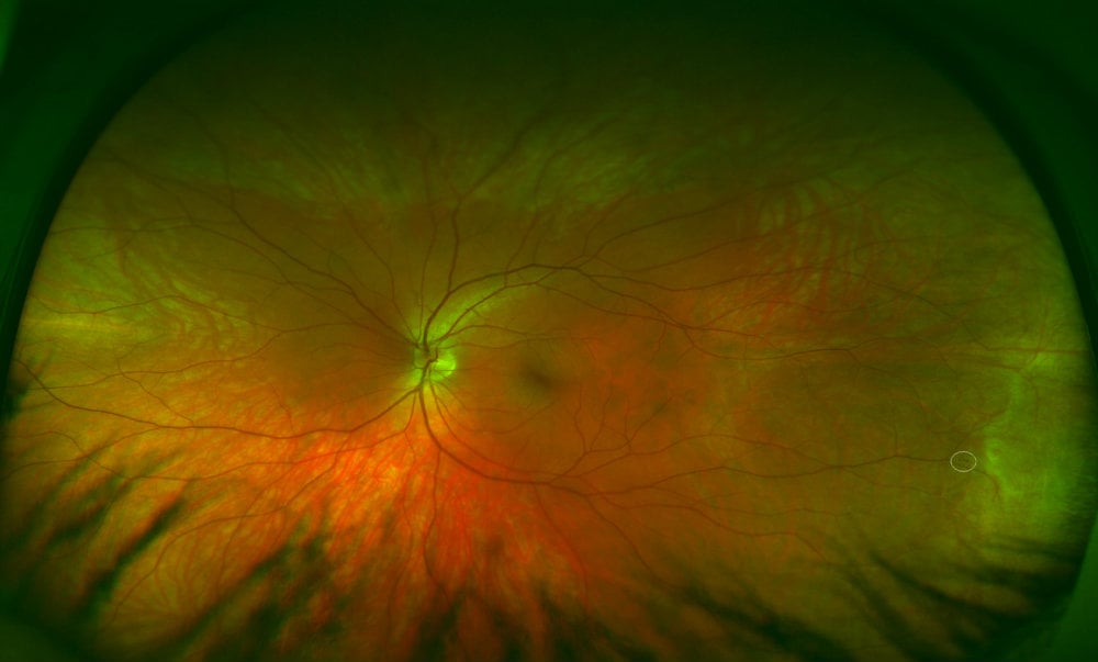
Far peripheral hemorrhages in a 32 year old not observed during traditional ophthalmoscopy.
In a press release, Roy Davis, CEO of Optos, shared that the study serves as a great example of the benefits of ultra-widefield imaging:
“We believe optomap image-assisted ophthalmoscopy represents an opportunity to improve pathology detection, the patient experience and to help a clinician efficiently target the area of the retina in need of further investigation. This targeted approach combined with the ability to electronically document pathologies can save valuable time and effort during examinations and raise the standard of care for patients.”
Kristen Brown, OD, FAAO, the principal study investigator, also affirms the many benefits of optomap, comparing it to smart technologies such as GPS:
“optomap images allow us to preview such a wide view of the retina before we perform our traditional exam, it’s like having a GPS of the retina. When you couple this with the benefits of digital management of the images such as zooming in to visualize key pathology, there are clear benefits in using this image-assisted technique.”
Practitioners interested in learning more about this study can read about it in contact us to request a consultation with an optomap representative.