Optos UWF autofluorescence helps classify AMD and predict progression
Examining the periphery with UWF autofluorescence (UWFAF) reveals patterns that suggest a new classification system for AMD and provide evidence of disease progression. A recent paper in Ophthalmology describes UWFAF imaging evaluations of patients with AMD (n=200) and no disease (n=19). The authors report that overall, 69% of eyes had peripheral AF abnormalities (86% with neovascular AMD, 73% with non-neovascular AMD, and 18% in eyes without AMD) and identify a strong correlation between observed AF patterns and the clinical features of the disease. They propose a classification system for AMD based on distinct AF patterns in the periphery and suggest that these patterns may be predictive of disease progression. The Optos 200Tx utilized in the study is the only imaging system available with the capability for UWFAF.
Tan CS, Heussen F, Sadda SR. Peripheral autofluorescence and clinical findings in neovascular and non-neovascularagre-related macular degeneration. Ophthalmology. 2013. [Epub ahead of print]
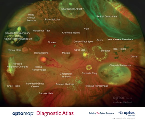
optomap® provides unprecedented visualization of retinal pathology in a single-capture digital image that can be readily annotated and shared. The new independently validated optomapDiagnostic Atlas™ displays pathologies that have been seen in our 200° view of the retina and is being supplied to doctors and staff to facilitate image interpretation and patient education.