Visualizing areas of retinal non-perfusion in patients with recalcitrant diabetic macular edema (DME) can provide important insights about the disease process and management. Results published in the American Journal of Ophthalmology suggest that utilizing ultra-widefield fluorescein angiography (UWFFA) to evaluate retinal vascular non-perfusion using the established ischemic index can identify DME that will be most unresponsive to therapy. This link between the extent of untreated retinal ischemia and the degree of recalcitrance provides further evidence that areas of non-perfusion produce biochemical mediators of disease progression, such as VEGF. The authors suggest that UWFFA could be useful in selecting patients for targeted retinal photocoagulation of untreated areas of non-perfusion and that employing the ischemic index could also help determine frequency of anti-VEGF therapy.
Patel RD, Messner LV, Teitelbaum B, Michele MA, Hariprasad SM. Characterization of ischemic index using ultra-widefield fluorescein angiograpy in patients with focal and diffuse recalcitrant diabetic macular edema. Am J Ophthalmol. 2013 [Epub ahead of print]
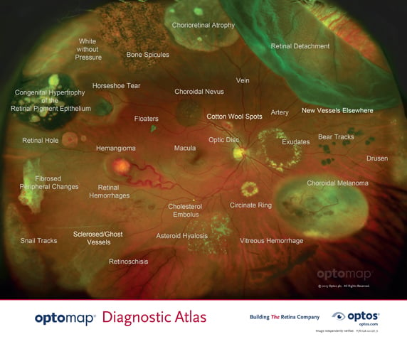
optomap® provides unprecedented visualization of retinal pathology in a single-capture digital image that can be readily annotated and shared. The new independently validated optomapDiagnostic Atlas™ displays pathologies that have been seen in our 200° view of the retina and is being supplied to doctors and staff to facilitate image interpretation and patient education.